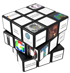
Our jobs at a glance
Towards novel hybrid imaging probes by chelator scaffolding
Concept: Molecular Imaging is an integral part of diagnostic and therapeutic algorithms rapidly evolving towards a novel standard for multimodality approaches. One way to combine different imaging modalities is based on the development of hybrid probes that provide a signal both in Positron Emission Tomography (PET) and Optical Imaging. This can be realized by combining a chelator for a radiometal (e.g. Gallium-68) with a fluorencent dye. Thereby, the information from radionuclide measurements can be seamlessly integrated in the results from optical detection e.g. within intraoperative procedures for image guided therapeutic applications.
The aim of this project is to develop novel multimodality imaging probes based on chelator scaffolding, expanding a novel concept (1) towards various oncological targets.
Approach: Within this PhD various cyclic chelator scaffold based probes will be synthetized targeting prostate cancer and neuroendocrine tumours. They will be characterized regarding radiolabelling properties, in vitro target binding and biodistribution, and finally investigated regarding their imaging properties.
Candidate: We search for a PhD student with a background in pharmacy, chemistry, biology or related fields, ideally with background in analytical techniques, biological assays or synthetic chemistry.
Co-supervision team:
Clemens Decristoforo (Radiopharmacist/PI): Researcher, Department of Nuclear Medicine, special expertise radiopharmaceutical development involving radiometals, peptides and siderophores and clinical translation.
Daniel Putzer (Radiologist/Co-PI): clinical scientist, Department of Radiology, special expertise in Image guided microinvasive therapies, Nuclear Medicine and image fusion
Ref: Summer D et al. Developing Targeted Hybrid Imaging Probes by Chelator Scaffolding. Bioconjug Chem. 2017 28(6):1722-1733.
Advanced fusion of 31P MRS and 1H MRI images and automatic brain tumor differentiation
Approach/methods/PhD:
With the existing imaging modalities, such as different MRI sequences or PET imaging, it is still difficult to distinguish between the different genetic mutations in glioblastoma multiforme (GBM) or differentiate between tumor progression and post-therapeutic changes. The aim of this project is to improve this with a combination of proton (1H) and phosphorous-based (31P) MR-spectroscopy (MRS) as well as other MRI and PET modalities
The aim of this project is to develop an automated image fusion method as well as a method, which can automatically differentiate tumor characteristics in GBM, with the help of affine transformations, interpolations, resampling techniques, variational methods and artificial neural networks. The data will be in the application of the machine learning and deep learning techniques for automatic differentiation.
Envisioned qualification of the candidates: student in applied mathematics / computer science / medicine/ engineering with first experience in medical imaging and/or application of machine learning methods.
Co-supervision team:
Markus Haltmeier (Mathematician/PI) applied mathematician, Department of Mathematics, special expertise on inverse problems, mathematical imaging, deep learning.
Astrid E. Grams (Radiologist/Co-PI): clinical scientist, Department of Radiology, special expertise in Neuroradiology, MR-spectroscopy, ultra high-field MRI, dual-energy CT, interventional radiology
CT/MR image fusion and automatic parameters estimation for TAVI procedure
Approach/methods/PhD:
The standard imaging work-up prior transcatheter aortic valve intervention (TAVI) is currently driven by a contrast-enhanced aortoiliacal computed tomography angiography (CTA). Fusion of a comprehensive cardiovascular MR image dataset with a non-contrast enhanced CT would combine the advantages of both modalities with immediate clinical applicability. Thus, in this project, we aim to develop software for fusing CT and MR images into one stacked image sequence (SIS) and to establish fully automatic data-driven parameter estimation techniques based on SIS. Mathematical image registration algorithms are developed which transform corresponding image regions of the two modalities into each other. For the automatic estimation of TAVI parameters, data-driven methods based on statistical learning and deep learning are developed, implemented and analyzed.
Envisioned qualification of the candidates is mathematics / computer science student with knowledge of medical imaging applications and good programming skills or medical student with additional expertise in mathematics or computer science.
Co-supervision team:
Agnes Mayr (Radiologist/PI): clinical scientist, Department of Radiology, special expertise in cardiovascular MRI.
Markus Haltmeier (Mathematician/Co-PI): applied mathematician, Department of Mathematics, special expertise on inverse problems, mathematical imaging, deep learning
Deep learning analysis of vascular imaging biomarker in vascular diseases
Approach/methods/PhD:
The accurate segmentation of cervical arteries from magnetic resonance (MR) or computer tomography (CT) images is a difficult challenge in radiology, but will allow for a quantitative analysis of arterial geometrical structure for the use in large cohort patient studies. Starting with preliminary work, we develop an automatic segmentation method with the help of appropriate affine transformations, interpolation, and resampling techniques. After manual delineation (supervised by neuroradiologist), the fusion method and the training and testing data will be created in supervision of the mathematician. This data will be used in the application of the machine learning techniques for the automatic differentiation.
Envisioned qualification of the candidates: medical student with additional expertise in computer science / applied mathematics / informatics with first experiences in medical imaging applications.
Co-supervision team:
Elke R. Gizewski (Radiologist/PI): clinical scientist, Department of Radiology, special expertise in Neuroradiology, functional, structural and metabolic MRI, ultra-high field MRI, neuro-interventions.
Lukas Neumann (Mathematician/Co-PI): Researcher, Department of Basic Sciences in Engineering, special expertise in inverse problems and image reconstruction methods, optimization and machine learning
Improvement of optical image quality by wavefront shaping
Approach/methods/PhD:
Imaging through tissues such as skin or brain at visible or near-infrared wavelengths is highly desired but impeded by severe light scattering. We will develop approaches for optical wavefront shaping in fluorescence microscopy using specifically designed fluorescent probes. The research aims at counteracting scattering, and consequently increasing the achievable imaging depth. Core technologies to be used include spatial light modulators and femto-second lasers for two-photon fluorescence excitation.
Envisioned qualification of the candidates: physics student with additional expertise in optical technologies and computer science.
Co-supervision team:
Monika Ritsch-Marte (Physicist/PI) in collaboration with Alexander Jesacher: physicists, Institute of Biomedical Physics (Biomedical Optics Lab)
Daniel Putzer (Radiologist/Co-PI) in collaboration with Clemens Decristoforo (Dept. of Nuclear Medicine)
Fast, non-rigid image fusion using deep learning for treatment evaluation after thermal ablation
Approach/methods/PhD:
In image-guided minimally-invasive percutaneous procedures, precise preoperative path planning is a key factor for the success of an intervention. This project will address automatizing trajectory planning of multiple needles, for efficient thermal ablation of abdominal tumors. The overall aim is to develop a (semi-) automatic framework facilitating the planning task of a surgeon for multiple needles, while also improving the quality of the planning and the overall safety for patients. A special focus will be on optimal geometric coverage of the intended tumor ablation volume, while minimizing the number of needles, and avoiding anatomical risk structures.
Envisioned qualification of the candidates: computer science or applied mathematics, with some experience in optimization and/or in medical applications.
Co-supervision team:
Matthias Harders (Computer Scientist/PI), Professor, Department of Computer Science, special expertise e.g. in physically-based simulation, surgical simulation/planning.
Reto Bale (Interventional Oncologist/Co-PI), clinical scientist, special expertise e.g. in stereotactic thermal ablation, percutaneous tumor and pain treatment
Multimodal magnetorelaxometry imaging of magnetic nonparticles
Approach/methods/PhD:
Magnetorelaxometry Imaging (MRXI) is a promising novel imaging approach enabling the non-invasive and quantitative imaging of magnetic nanoparticle (MNP) distributions, which represent a key element in a number of novel cancer therapies. In addition, MRXI has the potential to deliver information about the biological environment of the particles. In this respect, multimodal markers for magnetic, optical, and nuclear imaging have recently attracted great interest. In this project, MRXI of MNP distributions within biological environments will be investigated. The suitability and the imaging properties of optical and radio-labeled MNP in MRXI will be studied. Changes in relaxation properties and spatial distribution in different biological environments will be experimentally investigated by optical imaging and PET. Envisioned qualification of the candidates: student from the field of engineering, physics or pharmacy with first experience in (medical) imaging technology and/or magnetic/optical/radio-labelled tracers.
Co-supervision team:
Daniel Baumgarten (Biomedical Engineer/PI): Researcher, Institute of Electrical and Biomedical Engineering (UMIT), special expertise in medical imaging, diagnostic and therapeutic applications of magnetic nanoparticles.
Elke R. Gizewski (Radiologist/Co-PI): clinical scientist, Department of Radiology, special expertise in Neuroradiology, functional, structural and metabolic MRI, ultra-high field MRI, neuro-interventions
Augmenting microscopic navigated surgery with knowledge
Aims:
To create a feedback loop for quality control in navigation systems that current provide only positional information in endoscopic or microscopic. To identify 3D anatomical structures in the video stream in relation to structures segmented in preoperative radiologic imagery.
Approach and methods:
Machine learning approaches will be used to identify anatomical structures in live and recorded stereo video streams provided by a high-precision stereo-microscopic navigation system. A manually created preliminary atlas of paradigmatic ENT structures created from fused CT and MR data a will serve as ground truth to extract the according anatomic structures form the live video stream using computer vision and (un-)supervised machine learning approaches.
Envisioned qualification of the candidates: MSc in computer science, mathematics or physics with affinity to programming in C/C++, Qt, Python. Ideally you have experience in computer vision, machine learning, and seek a challenge to dive into the highly challenging medical technology domain.
Co-supervision team:
Wolfgang Freysinger (Medical Physicist/PI): Department of Otorhinolaryngology-Head & Neck Surgery, focused on navigation systems, visual user interfaces, application accuracy, all in clinics and laboratory. Extensive experience in clinical navigation.
Ute Ganswindt (Radiooncologist/Co-PI): clinical scientist, Department of Radiooncology, special expertise in IGRT, integration of functional imaging data (MRI/PET) into radiotherapy planning for improving safety and efficacy.
Stereoscopic X-ray, 3D surface imaging and CBCT for IGRT/SRS – cross modality evaluation
Aims:
To develop algorithms for the clinical use of surface imaging systems in comparison to CBCT/stereoscopic X-ray, in cases of targets underlying breathing motion to develop estimation models correlating internal vs. external image data.
Approach and methods: Systematic collection of inter-/intra-fractional 3D surface imaging data in clinical practice for several tumor entities/RT techniques. Data analyses: statistical determination of positioning variations, volumetric image data, mathematic modelling approaches.
Qualifications sought: MD with affinity to computer science and medical imaging/machine learning or student from the fields of computer science, mathematics, physics, engineering with affinity to medical imaging – seeking a challenge to dive into the highly challenging medical technology domain nearby clinical practice in radiation oncology.
Co-supervision team:
Ute Ganswindt (Radiooncologist/PI): clinical scientist, Department of Radiooncology, special expertise in IGRT, integration of functional imaging data (MRI/PET) into radiotherapy planning for improving safety and efficacy.
Daniel Baumgarten (Biomedical Engineer/Co-PI): Institute of Electrical and Biomedical Engineering (UMIT), focused on biological modeling and simulation with applications in medical imaging.
(Semi-)Automatic trajectory planning for multi-probe stereotactic thermal ablation of liver tumors
Approach/methods/PhD:
Thermal ablation is becoming a routine procedure for the treatment of liver tumors. Due to respiratory motion and the thermal ablation procedure itself, elastic deformation algorithms based on biomechanical representations of the relevant structures are required. This project will aim at new methods for non-rigid image fusion employing deep learning for treatment evaluation after thermal ablation, especially leveraging fast, deep learning approaches for physically-based simulations. Based on currently available methods in the involved research groups, the non-rigid deep learning approach will be developed, especially aiming at near real-time usage.
Envisioned qualification of the candidates: computer science or applied mathematics, with first experience in machine learning applications or medical science with additional expertise in computer science.
Co-supervision team:
Reto Bale (Interventional Oncologist/Co-PI), clinical scientist, special expertise e.g. in stereotactic thermal ablation, percutaneous tumor and pain treatment.
Matthias Harders (Computer Scientist/PI), technical lead, Professor, Department of Computer Science, special expertise e.g. in physically-based simulation, surgical simulation/planning
Novel Receptor-specific targeting in patients with advance neuroendocrine tumours
Approach, methods:
Different peptide analogues targeting the cholecystokinin-2 receptor and radiolabelled with lutetium-177 will be evaluated preclinically. Preclinical testing supervised by a radiopharmacist will include radiolabelling experiments, as well as in vitro / in vivo studies characterising the tumour targeting and therapeutic effect. Furthermore, receptor targeting using a conjugate labelled with a fluorescent dye is envisaged to investigate the potential for optical imaging. For one specific 177Lu-labelled minigastrin analogue, a clinically suitable radiopharmaceutical formulation will be developed and all the documents for a clinical trial application will prepared under the supervision of a clinical scientist. This preparatory work will be used for a first clinical trial evaluating the safety of administration as well as dosimetry aspects for peptide-receptor-radionuclide-therapy in patients with advanced neuroendocrine tumours by SPECT/CT imaging. Envisioned qualification of the candidate: pharmacist, biologist or comparable background in natural sciences, preferably with first experience in radiolabelling or fluorescent labelling / cell work / animal models / clinical studies.
Co-supervision team:
Elisabeth von Guggenberg (Radiopharmacist/PI): natural scientist, Department of Nuclear Medicine, with specific expertise in the preclinical and clinical evaluation of radiolabelled biomolecules.
Leonhard Gruber (Radiologist/Co-PI): clinical scientist, Department of Radiology, special expertise in data management, biostatistics, clinical imaging, and clinical translation of radiopharmaceuticals
Machine Learning for ENT Oncology
Aims:
This project aims at demonstrating the feasibility and clinical value of Radiomics, namely the use of novel machine learning approaches with limited amounts of data. Further, this project aims to provide a framework for clinicians in daily use with “diagnostic” capabilities concerning the malignancy of suspicious structures in CT imagery.
Approach and methods:
An open and closed source tools will be implemented and assessed on anonymized clinical tumor patient data. The thesis will build on Python and GPU-based machine learning, exploiting state-of-the-art libraries like Tensor Flow on a local or grid environment allowing for ample space of research and development. A desired outcome of the thesis could be a clinical trial with the developed tools. The work is grounded extensively on high-tech clinical infrastructures of both clinics involved.
Envisioned qualification of the candidates: MSc in computer science, mathematics or physics with affinity to programming in C/C++, Qt, Python, Linux, GPUs. Ideally you have experience in machine learning and seek a challenge to dive into the highly challenging medical technology domain.
Co-supervision team:
Wolfgang Freysinger (Medical Physicist/PI): Department of Otorhinolaryngology-Head & Neck Surgery, focused on navigation systems, visual user interfaces, application accuracy, all in clinics and laboratory. Extensive experience in clinical navigation.
Ute Ganswindt (Radiooncologist/Co-PI): clinical scientist, Department of Radiooncology, special expertise in IGRT, integration of functional imaging data (MRI/PET) into radiotherapy planning for improving safety and efficacy.
Deep learning- based strategy for scar pattern recognition on late gadolinium enhancement cardiac MRI
Approach/methods/PhD:
Late gadolinium enhancement (LGE) cardiac magnetic resonance imaging (CMR) is the current standard modality for myocardial scar assessment. The distribution pattern of LGE differs between ischemic and inflammatory injury with a typical subendocardial and subepicardial or midwall enhancement, respectively. While for the former its delineation from the blood pool may be challenging, for the latter the epicardial fat makes detection more difficult, which can lead to a complex and observer-dependent diagnosis. In this project a deep-learning based strategy for segmentation of the myocardial borders and LGE pattern recognition are developed aimed to facilitate objective CMR diagnosis. Different neural network based methods will be developed, numerically realized, evaluated and extended. Networks will be developed taking into account physical and clinical constraints and combined with methods of variational imaging. Devolved methodologies will subsequently be extended to related clinical research problems.
Envisioned qualification of the candidates is mathematics / computer science student with knowledge of medical imaging applications and good programming skills.
Co-supervision team:
Markus Haltmeier (Mathematician/PI): applied mathematician, Department of Mathematics, special expertise on inverse problems, mathematical imaging, deep learning.
Agnes Mayr (Radiologist/PI): clinical scientist, Department of Radiology, special expertise in cardiovascular MRI
Feature supported deep learning for image guided diagnosis
Approach/methods/PhD:
Establish deep learning methods for joint image reconstruction, enhancement, analysis and interpretation, include image features stabilizing diagnostics and increasing interpretability.
Project description:
This project is naturally related to PhD4 on deep learning of vascular imaging biomarkers. Contrary to the approach followed in PhD4, here we aim at improving image reconstruction and image analysis in the same step. We expect that by using joint loss functions and hybrid network architectures significant improvements can be made. However the risk associated with misinterpretation may be higher and thus such an approach needs to be evaluated even more thoroughly before it can be implemented in clinical environments. Employing generative adversarial networks we expect that parts of the training can still be done unsupervised. A profound mathematical understanding of the problem will be necessary to gain a clear picture of the benefits and possible risks of such a strategy.
Envisioned qualification of the candidates: student in applied mathematics / computer science / medicine/ engineering with first experience in medical imaging and/or application of machine learning methods.
Co-supervision team:
Lukas Neumann (Mathematician/PI): Researcher, Department of Basic Sciences in Engineering, special expertise in inverse problems and image reconstruction methods, optimization and machine learning.
Elke R. Gizewski (Radiologist/Co-PI): clinical scientist, Department of Radiology, special expertise in Neuroradiology, functional, structural and metabolic MRI, ultra-high field MRI, neuro-interventions.
Stephanie Mangesius (Radiologist/Co-PI): clinical scientist, Department of Radiology, special expertise in Neuroradiology, functional, structural and metabolic MRI, ultra-high field MRI, quantitative imaging and AI
Multinuclear 23Na and 1H quantitative MRI in brain tumors and metastases
Aims:
Sodium (23Na) is the second most abundant MRI active nucleus and plays an important role in the human brain’s metabolism. In several pathologies, such as cancer, a disturbed balance of the intra- and extracellular sodium concentration is present. In brain tumors, an increase was observed in total sodium concentration attributed to a disturbed cellular energy metabolism. Therefore, 23Na-MRI could provide valuable information in addition to 1H-MRI aiming for a more precise characterization of pathological changes in brain tumors and metastases prior and upon treatment. The aim of this project is to develop a hybrid imaging approach by combining 23Na and 1H quantitative MRI in a clinical setting with patients with brain tumors or brain metastases. One major focus will be the quantification of the multi-exponential 23Na and 1H effective transversal relaxation rate R2. This should be achieved by adapting existing MRI sequences to assess multi-echo 23Na and 1H images and by developing an image analysis method to separate slow and fast components of the total R2 signal decay for both 23Na and 1H. The different R2* components can than further be assigned to different microstructural tissue compartments, such as the intra- and extracellular sodium and water components. By applying the developed approach to the patients will allow to assess pathological differences in multi-compartmental 23Na and 1H relaxation in different brain tumors. For patients with metastases, this approach will allow the monitoring of treatment-induced microstructural changes. As 23Na and 1H imaging relies on different MR coils and MR sequences, image registration will be an important aspect. Therefore, this PhD project covers tasks from adapting and testing MR sequences, quantification of MR parameters, classical (non-AI) image analysis and the work in an interdisciplinary patient study.
Envisioned qualification of the candidates: Biomedical engineering or physics with an experience in medical image analysis and MRI, advanced programming skills and an interest in basic medicine/neuroscience.
Co-supervision team:
Christoph Birkl (MR physicist/PI): Researcher, Department of Neuroradiology, special expertise in MR physics, quantitative imaging, medical image analysis and biophysics.
Julian Mangesius (Radiooncologist/CO-PI): clinical scientist, Department of Radiooncology, special expertise in neurooncology, image-guided radiation therapy and stereotactic radiation therapy
Daniel Baumgarten (Biomedical Engineer/CO-PI): Researcher, Institute of Electrical and Biomedical Engineering (UMIT), special expertise in medical imaging, biological modeling and simulation
Ute Ganswindt (Radiooncologist/Co-PI): clinical scientist, Department of Radiooncology, special expertise in image-guided radiation therapy, integration of imaging data into radiotherapy planning for improving safety and efficacy

